
As I mentioned in my previous post (https://livingwithfh.blogspot.com/2023/09/old-test-new-reaction.html), my cardiologist and the vascular surgeon ordered a CT scan with contrast of my abdominal aorta. This test was done in 2021 and it showed several areas of atherosclerosis of basically every branch of my abdominal aorta.
Now, two years later, it shows the same areas of problems - some of the areas look stable (yay!), and some are slightly worse, or show new developments. For example, I have developed “collaterals”, which means additional (new) branches of the abdominal aorta that naturally grew to bypass the areas that are stenotic. So, the fact that the collaterals are there at all, to help with the proper irrigation of the gut area, is good, but the fact that they had to form at all says the original branches don’t provide proper circulation to the abdomen.
In addition to the stenoses caused by the calcified plaque, because this is me, and I am not easy, I also have a congenital defect of my celiac abdominal aorta called “median arcuate ligament compression“ (or MAL compression) which is rare condition in which the median arcuate ligament compresses the celiac artery which impairs blood flow to the liver, stomach and other organs. The median arcuate ligament is the muscle and fibrous structure that wraps around the aorta at the diaphragmatic opening. So, this compression in addition to the atherosclerotic celiac artery causes a reduced flow to my abdomen.
Here are the test results of the two scans, over the past two years:
The December 2021 abdominal CT scan results show the following:
Small caliber abdominal aorta with extensive soft and calcified plaque
Narrowing of the infrarenal abdominal aorta
Two areas of moderate to severe stenosis in the proximal celiac artery and atherosclerotic plaque at the ostium and compression from the median arcuate ligament (MAL) with mild poststenotic dilatation.
Mild stenosis at the origin of the small mesenteric artery.
The September 2023 abdominal CT scan results show the following:
Stable: Severe calcified and noncalcified atherosclerosis of the entire abdominal aorta which is overall small in caliber, unchanged.
Stable: There is mild to moderate relative narrowing of the infrarenal abdominal aorta at the inferior mesenteric artery, also unchanged.
Slightly worse: Severe stenosis and angulation of the proximal celiac artery likely owing to a combination of atherosclerotic plaque and the median arcuate ligament (MAL) compression. (previously: “moderate to severe”)
Slightly worse: Persistent moderate stenosis of the proximal superior mesenteric artery (previously: “mild”)
New (good and bad):
The presence of arterial collaterals between the celiac-superior mesenteric artery and to a lesser extent the superior - inferior mesenteric arteries territories suggest the presence of long-standing, hemodynamically significant stenosis in at least one of these vascular territories.
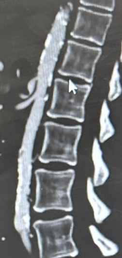
In this scan, the white part to the left of my spine is the calcified aorta. The smaller vessels springing from it are my celiac (the one in the shape of a backwards L which shows the MAL compression) and the mesenteric arteries.
Although the tests seem virtually unchanged, I have new symptoms that could be related to the various stenoses in my abdominal aorta. Some of these symptoms are a fairly consistent upper-abdominal pain, which is worse after I eat; the feeling of being “full” even when I first wake up in the morning and I have an empty stomach; occasional nausea with even mild exercise, softer and more often stools.
When I went in to the vascular surgeon this past week to review these results, the surgeon got called into emergency surgery and was not able to make our appointment. I spoke, instead, to one of the PAs (not his own PA, who I knew and who knew my case, but a new one), and having just seen me for the first time, he said that the new symptoms worry him and he will need to chat with the doctor and see what, if anything, needs to be done next. But he also said that in his opinion, the results are not much different than the ones from two years ago, so the symptoms might not be related to the stenoses.
I also mentioned that I have been examined by a gastro-enterologist earlier this year, to ensure none of these new symptoms are GI related and they are not, according to those tests. He said he will follow up with them and then he and the vascular surgeon will follow up with a plan, if there is anything to be done differently. At the very least, he suggested that I’d move the scans to every 6 months rather than every year to keep a closer eye on the abdominal aorta.
Because the risk of too much radiation during a year’s time is there, he suggested I’d alternate between an ultrasound and a CT scan, every 6 months. Although I dread all these different tests (I have another one for my carotid arteries and another one for my heart - both yearly), I know that keeping a close eye on these conditions is really the best way to catch some huge change in time, before having an event. If my annual heart echos taught me anything before I had my open-heart surgery, they taught me this.